Login
Welcome back! Please enter your details.
or
Don't have an account? Register here
Create Account
Join MedMentorEdu and start your medical journey.
or
Already have an account? Login here
Enhance your knowledge with our comprehensive guide and curated study materials.
Study of microorganisms (not visible to naked eye)
Includes bacteria, viruses, fungi, parasites, prions
Focus: structure, function, genetics, pathogenicity
Medical microbiology → disease causation, diagnosis
Industrial microbiology → antibiotics, vaccines
Environmental microbiology → nutrient cycles
Food microbiology → fermentation, spoilage
Molecular microbiology → genetic engineering
Bacteriology → bacteria
Virology → viruses
Mycology → fungi
Parasitology → protozoa, helminths
Immunology → host defense mechanisms
Antonie van Leeuwenhoek
First to observe microorganisms
“Father of Microbiology”
Louis Pasteur
Disproved spontaneous generation
Fermentation and pasteurization
Vaccine development
Robert Koch
Established germ theory of disease
Formulated Koch’s postulates
Organism present in diseased host
↓
Isolate and grow in pure culture
↓
Inoculate into healthy host → disease reproduced
↓
Re-isolate same organism
Identification of pathogens
Basis of diagnosis, treatment, prevention
Development of vaccines, antibiotics
Understanding epidemiology
Normal flora
Present in body without causing disease
Protective role (colonization resistance)
Pathogens
Cause disease
Can be:
Primary
Opportunistic
Infection
Entry and multiplication of microorganisms
Disease
Clinical manifestation due to infection
Primary infection → initial infection
Secondary infection → follows primary infection
Opportunistic infection → occurs in immunocompromised host
Subclinical infection → no symptoms
Natural habitat of organism
Types:
Human reservoir
Animal reservoir
Environmental reservoir (soil, water)
Direct
Person-to-person contact
Droplet spread
Indirect
Fomites
Airborne
Vector-borne
Mosquito, ticks
Sterilization
Complete destruction of all microorganisms including spores
Disinfection
Removal of pathogenic organisms (not spores)
Culture
Growth of microbes in artificial media
Media
Nutrient substance for growth
Pathogen
Disease-causing organism
Opportunist
Causes disease in weakened host
| Branch | Organism Studied |
|---|---|
| Bacteriology | Bacteria |
| Virology | Viruses |
| Mycology | Fungi |
| Parasitology | Protozoa, helminths |
| Immunology | Immune system |
| Scientist | Contribution |
|---|---|
| Leeuwenhoek | First microscope observations |
| Pasteur | Germ theory, vaccines |
| Koch | Koch’s postulates |
| Type | Description |
|---|---|
| Primary | Initial infection |
| Secondary | Follows primary |
| Opportunistic | In immunocompromised |
| Subclinical | No symptoms |
| Mode | Examples |
|---|---|
| Direct | Contact, droplets |
| Indirect | Fomites, airborne |
| Vector-borne | Mosquito, ticks |


Description (Exam points):
Visible colonies on agar
Each colony = clonal population
Important features:
Shape
Margin
Elevation
Pigmentation
Commensalism → Mutualism → Parasitism
             ↓
        Disease (pathogenicity)
Infectious agent
↓
Reservoir
↓
Portal of exit
↓
Mode of transmission
↓
Portal of entry
↓
Susceptible host
All living organisms are composed of cells
Cell is the basic structural and functional unit
Cells arise from pre-existing cells
Microorganisms follow same fundamental cellular principles
Prokaryotes
No true nucleus
No membrane-bound organelles
DNA in nucleoid
Smaller size (1–5 µm)
Example: Bacteria
Eukaryotes
True nucleus present
Membrane-bound organelles
Larger size (10–100 µm)
Example: Fungi, protozoa
Molecular → DNA, RNA, proteins
Cellular → individual cells
Tissue → group of cells
Organ → functional unit
Organism → complete living entity
Genetic information flow:
DNA → replication
DNA → RNA (transcription)
RNA → Protein (translation)
DNA
↓ (Transcription)
RNA
↓ (Translation)
Protein
Genotype
Genetic constitution of organism
Phenotype
Observable characteristics
Influenced by environment + genotype
Mutation
Permanent change in DNA sequence
Types:
Point mutation
Frameshift mutation
Leads to:
Variation
Drug resistance
Virulence changes
Microbes evolve through:
Mutation
Genetic recombination
Natural selection
Survival of organisms with advantageous traits
Transfer of genetic material between organisms
Types:
Transformation
Uptake of naked DNA
Transduction
Transfer via bacteriophages
Conjugation
Transfer via sex pili
Genetic mutation
Gene transfer
Enzyme production
Biofilm formation
Antimicrobial resistance
Commensalism
One benefits, other unaffected
Mutualism
Both benefit
Parasitism
One benefits, host harmed
Pathogenicity
Ability to cause disease
Virulence
Degree of severity of disease
Innate immunity
First line defense
Non-specific
Adaptive immunity
Specific response
Memory present
| Feature | Prokaryotes | Eukaryotes |
|---|---|---|
| Nucleus | Absent | Present |
| Organelles | Absent | Present |
| DNA | Circular | Linear |
| Size | Small | Large |
| Ribosomes | 70S | 80S |
| Type | Description |
|---|---|
| Commensalism | One benefits, other unaffected |
| Mutualism | Both benefit |
| Parasitism | Host harmed |
| Feature | Pathogenicity | Virulence |
|---|---|---|
| Definition | Ability to cause disease | Severity of disease |
| Nature | Qualitative | Quantitative |
| Type | Mechanism |
|---|---|
| Transformation | Naked DNA uptake |
| Transduction | Phage-mediated |
| Conjugation | Sex pili transfer |

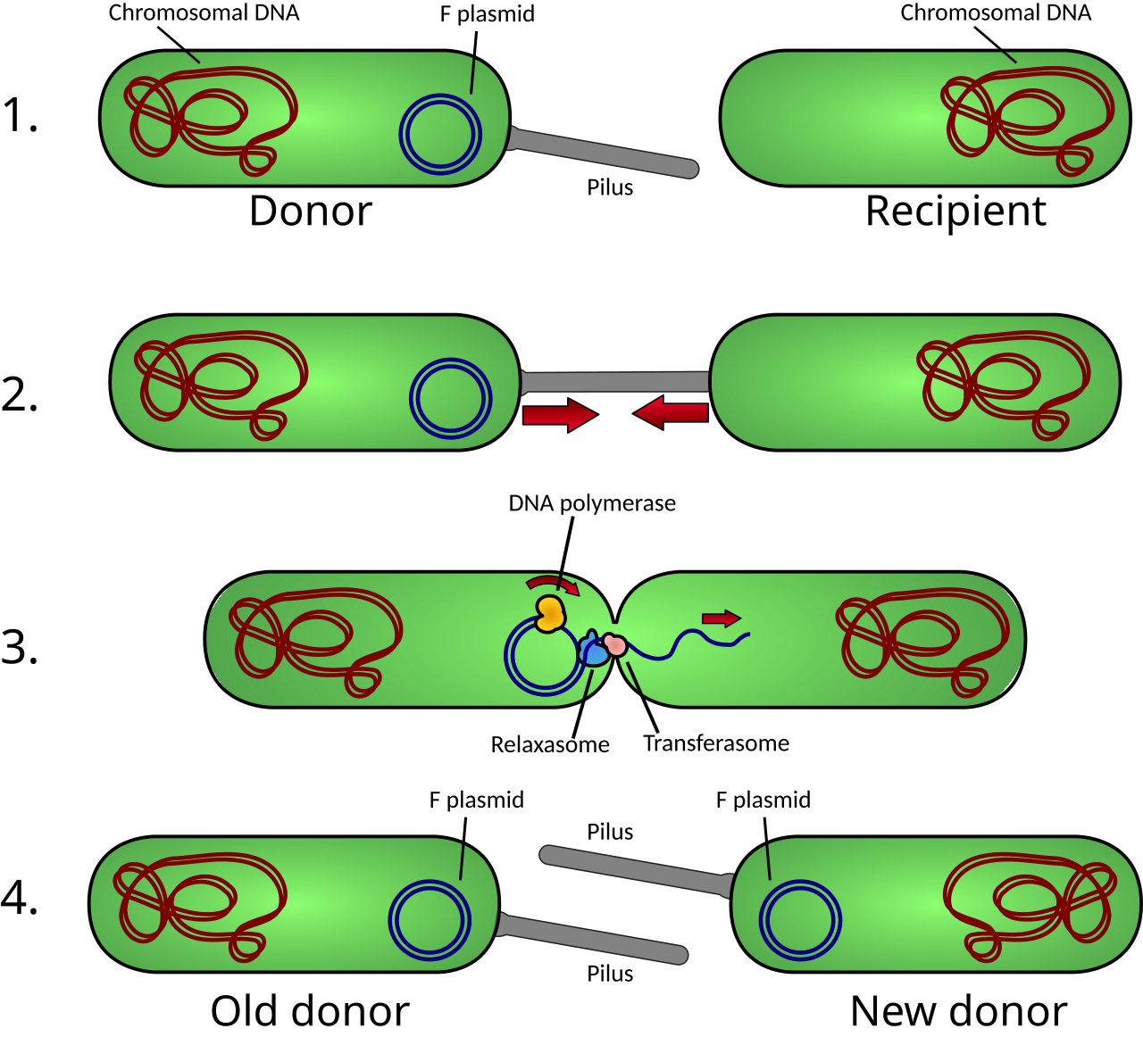
Description (Exam points):
Two bacteria connected by sex pilus
DNA transfer from donor (F+) to recipient (F−)
Important mechanism of antibiotic resistance spread
Mutualism → Commensalism → Parasitism
                        ↓
                   Disease
Transformation → Naked DNA uptake
Transduction → Phage-mediated transfer
Conjugation → Sex pilus DNA transfer
Mutation
↓
Genetic variation
↓
Natural selection
↓
Adaptation
↓
Evolution
Viruses are obligate intracellular parasites
Contain single type of nucleic acid (DNA or RNA)
Lack cellular organelles
Cannot grow on artificial media
Replicate only inside living cells
Capsid
Protein coat
Made of capsomeres
Envelope
Lipid layer derived from host
Contains glycoprotein spikes
Nucleic Acid
DNA or RNA
Single-stranded or double-stranded
Icosahedral
Spherical appearance
Example: Adenovirus
Helical
Rod-shaped
Example: Rabies virus
Complex
Combination structure
Example: Bacteriophage
Based on:
Nucleic acid type
Presence of envelope
Symmetry
Replication strategy
Based on type of nucleic acid and replication method
| Group | Genome Type |
|---|---|
| I | dsDNA |
| II | ssDNA |
| III | dsRNA |
| IV | ssRNA (+) |
| V | ssRNA (−) |
| VI | RNA with reverse transcriptase |
| VII | DNA with reverse transcriptase |
Steps:
Attachment
Penetration
Uncoating
Replication
Assembly
Release
Attachment
↓
Penetration
↓
Uncoating
↓
Replication
↓
Assembly
↓
Release
Lytic cycle
Virus replicates → host cell destroyed
Lysogenic cycle
Viral DNA integrates into host genome
Remains latent
Phases:
Latent phase
Eclipse phase
Burst phase
Structural changes in infected cells
Examples:
Cell lysis
Syncytium formation
Inclusion bodies
Aggregates of viral particles
Examples:
Negri bodies → Rabies
Owl’s eye inclusion → CMV
Viruses infecting bacteria
Have head, tail, tail fibers
Can undergo lytic or lysogenic cycle
DNA viruses → replicate in nucleus
RNA viruses → replicate in cytoplasm
RNA viruses mutate rapidly
Viruses causing cancer
Examples:
HPV
EBV
HBV
| Feature | DNA Viruses | RNA Viruses |
|---|---|---|
| Genome | DNA | RNA |
| Replication site | Nucleus | Cytoplasm |
| Mutation rate | Low | High |
| Feature | Enveloped | Non-Enveloped |
|---|---|---|
| Outer layer | Lipid envelope | Absent |
| Stability | Fragile | Resistant |
| Transmission | Direct contact | Fomites |
| Stage | Description |
|---|---|
| Attachment | Virus binds to host |
| Penetration | Entry into cell |
| Uncoating | Release of genome |
| Replication | Genome synthesis |
| Assembly | Formation of virions |
| Release | Exit from cell |
| Type | Shape | Example |
|---|---|---|
| Icosahedral | Spherical | Adenovirus |
| Helical | Rod-shaped | Rabies |
| Complex | Irregular | Bacteriophage |
| Feature | Lytic | Lysogenic |
|---|---|---|
| Host cell | Destroyed | Survives |
| Viral DNA | Independent | Integrated |
| Outcome | Rapid replication | Latency |


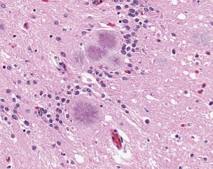
Description (Exam points):
Eosinophilic inclusions in neurons
Seen in rabies infection
Diagnostic significance


Description (Exam points):
Cell rounding and detachment
Syncytium formation
Cell lysis


Description (Exam points):
Clear zones in bacterial lawn
Each plaque = infection by single virus
Used for viral quantification
Capsid
↓
Nucleic acid
↓
Envelope (optional with spikes)
Lytic: Entry → Replication → Cell lysis

Lysogenic: Entry → Integration → Latency → Activation → Lysis
Latent phase → Eclipse phase → Burst phase
Head (DNA)
↓
Neck
↓
Tail
↓
Tail fibers
Prions are proteinaceous infectious particles
Lack nucleic acid (no DNA or RNA)
Cause neurodegenerative diseases
Composed of misfolded prion protein (PrPsc)
Derived from normal cellular protein (PrPc)
Highly resistant to:
Heat
Radiation
Disinfectants
Normal protein (PrPc) converted to abnormal (PrPsc)
Misfolded protein induces further misfolding
Normal PrPc
↓
Interaction with PrPsc
↓
Misfolding
↓
Accumulation of PrPsc
↓
Neuronal damage
Creutzfeldt-Jakob Disease (CJD)
Kuru
Bovine Spongiform Encephalopathy (BSE)
Transmission between species is limited
Due to differences in PrP structure
Barrier can be crossed (e.g., BSE → humans)
Spongiform degeneration
Vacuolation in brain tissue
Neuronal loss
Gliosis
No inflammatory response
No nucleic acid
Resistant to conventional sterilization
Long incubation period
Not detected by routine immune response
| Feature | Prions | Viruses |
|---|---|---|
| Genetic material | Absent | DNA/RNA present |
| Structure | Protein only | Nucleic acid + protein |
| Replication | Protein misfolding | Host cell machinery |
| Resistance | Highly resistant | Less resistant |
| Type | Example |
|---|---|
| Sporadic | CJD |
| Familial | Genetic CJD |
| Acquired | Kuru, BSE |


Description (Exam points):
Multiple vacuoles in gray matter
“Sponge-like” appearance
Neuronal loss without inflammation
Prion entry
↓
Conversion of PrPc → PrPsc
↓
Accumulation in brain
↓
Neuronal damage
↓
Spongiform degeneration
Unicellular organisms without true nucleus
DNA present as nucleoid (circular DNA)
Lack membrane-bound organelles
Size: 1–5 µm
Multiply by binary fission
Cell wall usually present
Provides shape and rigidity
Protects against osmotic lysis
Polymer of:
N-acetylglucosamine (NAG)
N-acetylmuramic acid (NAM)
Cross-linked by peptide bridges
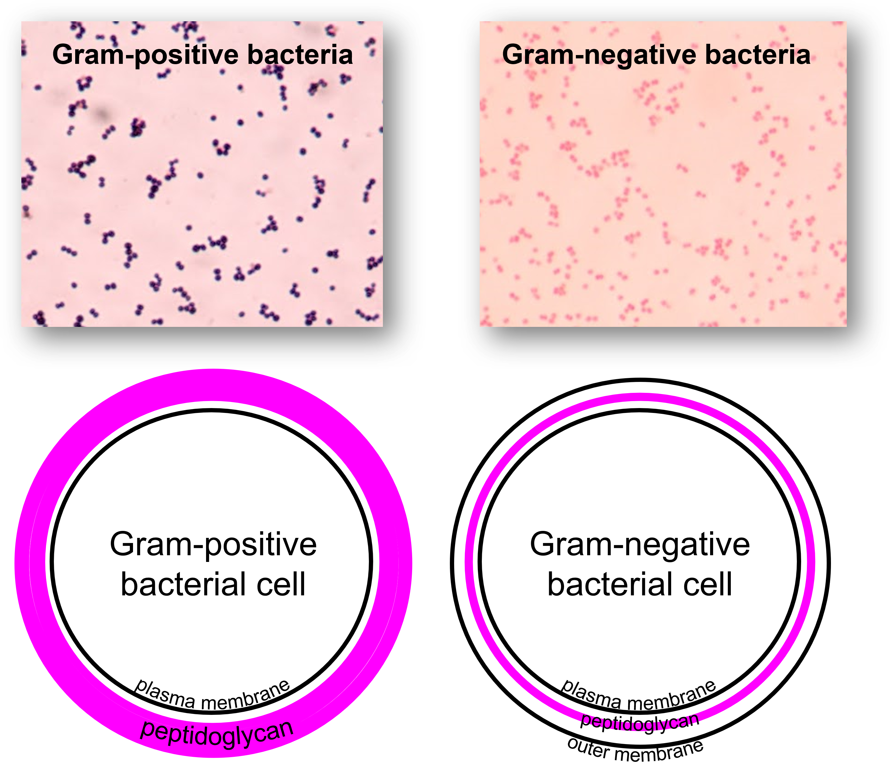
Present in Gram-positive bacteria
Functions:
Cell wall stability
Antigenic properties
Present in Gram-negative bacteria
Components:
Lipid A → endotoxin
Core polysaccharide
O antigen
Phospholipid bilayer
Site of:
Respiration
Enzyme activity
Transport
Contains enzymes, nutrients
No organelles
Circular double-stranded DNA
No nuclear membrane
70S type
Site of protein synthesis
Polysaccharide layer
Functions:
Antiphagocytic
Virulence factor
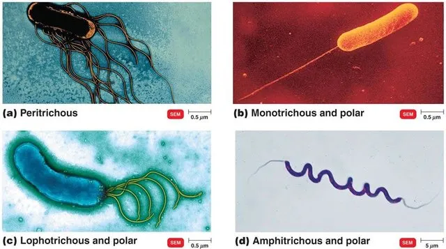
Motility structures
Composed of flagellin
Types:
Monotrichous
Lophotrichous
Amphitrichous
Peritrichous
Hair-like structures
Functions:
Adhesion
Conjugation (sex pili)
Steps:
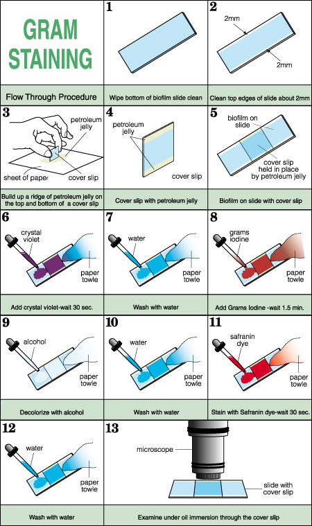
Crystal violet (primary stain)
Iodine (mordant)
Alcohol (decolorizer)
Safranin (counterstain)
Result:
Gram-positive → purple
Gram-negative → pink
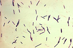
Cocci → spherical
Bacilli → rod-shaped
Spiral forms → spirilla, spirochetes
Arrangements:
Chains (strepto-)
Clusters (staphylo-)
Pairs (diplo-)
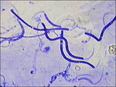
Flagellar motility
Gliding motility
Twitching motility
Binary fission
Rapid multiplication
Generation time varies
Phases:
Lag phase
Adaptation
No division
Log phase
Rapid multiplication
Maximum metabolic activity
Stationary phase
Nutrient depletion
Growth = death
Decline phase
Cell death predominates
Energy production via:
Aerobic respiration
Anaerobic respiration
Fermentation
Autotrophs → use CO₂
Heterotrophs → use organic compounds
Subtypes:
Saprophytes
Parasites
Obligate aerobes
Obligate anaerobes
Facultative anaerobes
Microaerophilic
Formation of endospores under adverse conditions
Highly resistant to:
Heat
Chemicals
Example: Bacillus, Clostridium
Plasmids
Extrachromosomal DNA
Carry resistance genes
Episomes
Can integrate into host DNA
| Feature | Gram-Positive | Gram-Negative |
|---|---|---|
| Cell wall | Thick peptidoglycan | Thin peptidoglycan |
| Teichoic acid | Present | Absent |
| LPS | Absent | Present |
| Staining | Purple | Pink |
| Structure | Function |
|---|---|
| Cell wall | Shape, protection |
| Capsule | Virulence, protection |
| Flagella | Motility |
| Pili | Adhesion, conjugation |
| Ribosomes | Protein synthesis |
| Shape | Example |
|---|---|
| Cocci | Staphylococcus |
| Bacilli | E. coli |
| Spiral | Treponema |
| Type | Example |
|---|---|
| Obligate aerobe | Mycobacterium |
| Obligate anaerobe | Clostridium |
| Facultative anaerobe | E. coli |
| Microaerophilic | Helicobacter |
| Type | Description |
|---|---|
| Autotroph | Uses CO₂ |
| Heterotroph | Uses organic nutrients |
| Saprophyte | Dead matter |
| Parasite | Living host |
| Phase | Feature |
|---|---|
| Lag | Adaptation |
| Log | Rapid growth |
| Stationary | No net growth |
| Decline | Death phase |
| Component | Function |
|---|---|
| Peptidoglycan | Strength |
| Teichoic acid | Stability |
| LPS | Endotoxin |

Description (Exam points):
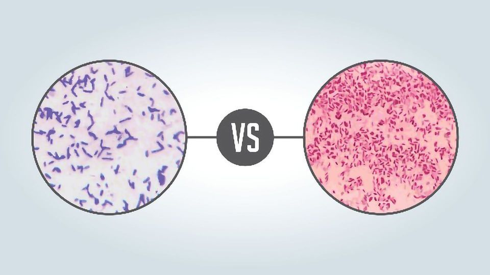
Gram-positive → purple (thick peptidoglycan)
Gram-negative → pink (thin wall + LPS)
Key diagnostic method
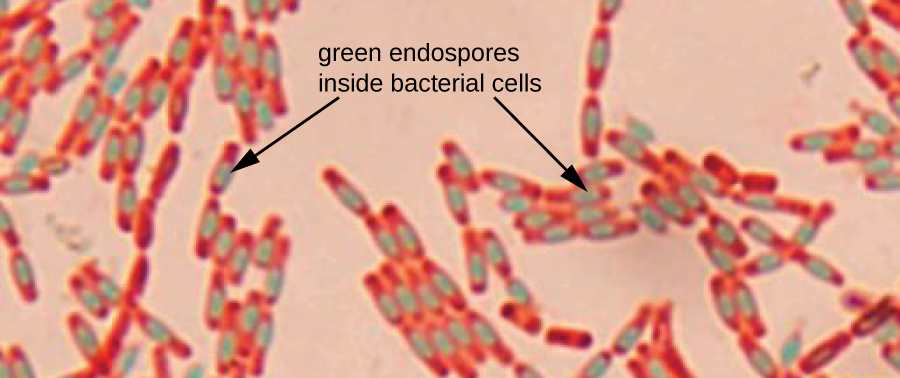

Description (Exam points):
Endospores appear green
Vegetative cells red
Indicates resistant bacteria
Capsule
↓
Cell wall
↓
Cell membrane
↓
Cytoplasm
↓
Nucleoid
↓
Ribosomes
Crystal violet → Iodine complex
↓
Alcohol wash
↓
Gram (+): retains color
Gram (−): loses color
↓
Safranin counterstain
Lag → Log → Stationary → Decline
Vegetative cell
↓
Spore formation
↓
Dormant spore
↓
Germination → active cell


Description (Exam points):
Purple spherical bacteria
Arrangement:
Clusters → Staphylococcus
Chains → Streptococcus
Thick peptidoglycan


Description (Exam points):
Pink rod-shaped bacteria
Thin cell wall + outer membrane
Example: E. coli


Description (Exam points):
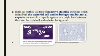
Clear halo around bacteria
Capsule unstained
Important virulence factor
Description (Exam points):
Green spores within red cells
Highly resistant structures
Seen in Bacillus, Clostridium
Description (Exam points):
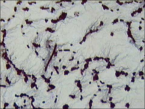
Thin filamentous structures
Helps identify motility pattern
Special staining required

Description (Exam points):
Crystal violet → Iodine → Alcohol → Safranin
Differentiates Gram + and Gram −


Description (Exam points):
Red bacilli on blue background
Mycolic acid in cell wall
Example: Mycobacterium tuberculosis
Capsule
↓
Cell wall
↓
Cell membrane
↓
Cytoplasm
↓
Nucleoid
↓
Ribosomes
Crystal violet → Iodine complex
↓
Alcohol wash
↓
Gram (+): retains color
Gram (−): decolorized
↓
Safranin counterstain
Protists are unicellular eukaryotic organisms
Include protozoa, algae, slime molds
Possess true nucleus and organelles
Protozoa → animal-like protists
Algae → photosynthetic
Slime molds → fungus-like
Eukaryotic structure
Motility by:
Pseudopodia
Flagella
Cilia
Reproduction:
Asexual (binary fission)
Sexual (some species)
Habitat: water, soil, host organisms
Amoebae → pseudopodia (e.g., Entamoeba)
Flagellates → flagella (e.g., Giardia)
Ciliates → cilia (e.g., Balantidium)
Sporozoa → non-motile (e.g., Plasmodium)
Trophozoite
Active, feeding stage
Motile
Sensitive to environment
Cyst
Dormant, infective stage
Resistant to harsh conditions
Transmission form
Involve:
Trophozoite stage
Cyst stage
May require:
Single host
Multiple hosts (e.g., malaria)
Fecal-oral route
Vector-borne (mosquito in malaria)
Contaminated food and water
Pathogenic
Cause disease
Example: Entamoeba histolytica
Non-pathogenic
Harmless commensals
Example: Entamoeba coli
Cause major diseases:
Amoebiasis
Giardiasis
Malaria
Important in public health
| Group | Mode of Movement | Example |
|---|---|---|
| Amoebae | Pseudopodia | Entamoeba |
| Flagellates | Flagella | Giardia |
| Ciliates | Cilia | Balantidium |
| Sporozoa | Non-motile | Plasmodium |
| Organism | Disease |
|---|---|
| Entamoeba histolytica | Amoebiasis |
| Giardia lamblia | Giardiasis |
| Plasmodium | Malaria |
| Feature | Trophozoite | Cyst |
|---|---|---|
| Activity | Active | Dormant |
| Motility | Present | Absent |
| Resistance | Low | High |
| Role | Feeding stage | Infective stage |
| Type | Example |
|---|---|
| Amoebae | Entamoeba histolytica |
| Flagellates | Giardia lamblia |
| Sporozoa | Plasmodium |

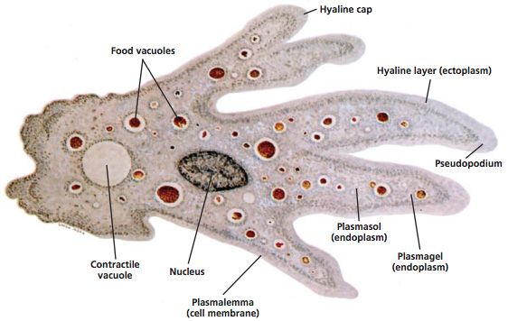

Description (Exam points):
Irregular shape with pseudopodia
Central nucleus
May contain ingested RBCs

![]()
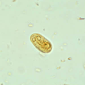
Description (Exam points):
Pear-shaped organism
Two nuclei (“face-like appearance”)
Flagella present
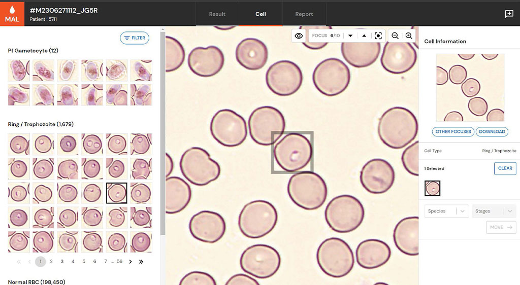

Description (Exam points):
Ring forms inside RBC
Schizonts with multiple nuclei
Gametocytes (banana-shaped in falciparum)
Cyst (infective stage)
↓
Ingestion
↓
Trophozoite (active stage)
↓
Multiplication
↓
Encystation
↓
Excretion of cyst
Ingestion of cyst
↓
Excystation in intestine
↓
Trophozoite formation
↓
Multiplication
↓
Encystation
↓
Cyst passed in stool
Amoebae → Pseudopodia
Flagellates → Flagella
Ciliates → Cilia
Sporozoa → Non-motile
Get the full PDF version of this chapter.